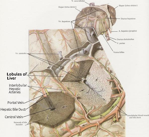
The lobules of liver constitute the histologic and fundamental units of this glandular organ. Polygonal in shape, each one of them measures between 1 and 2 mm in size. They are surrounded by an interlobular septum, which is composed of fibrous connective tissue. The lobules exhibit a distinctive radial pattern of hepatocyte rows, which converge towards a central vein.
Histologic Description
The lobules of liver consist of cells called hepatocytes, which are the parenchymal (functional) cells of this organ. The hepatocytes are arranged in a radial pattern. In the center of each lobule, there is a central vein. There is also a portal canal in each corner of a lobule. Each portal canal contains in turn an interlobular hepatic artery, a portal vein, and a hepatic vile duct. The portal vein is a small branch of the hepatic portal vein, which brings in nutrient-rich blood from the superior mesenteric vein to be metabolized by the hepatocytes.
The interlobular capillaries enter the lobule, in which they are continuous with the sinusoids lying between the hepatic plates. These sinusoids contain mixed arterial and venous blood, which drain into the central vein. This central vein in turn drains into the collecting sublobular veins, which empty the deoxygenated blood into the right, middle, and left hepatic vein.
Among the hepatocytes contained within a lobule, there are the biliary canaliculi, which drain into the bile ductules (ductuli biliferi). The latter get together and unite outside the lobule of liver to form the interlobular bile ducts. Furthermore, the interlobular bile ducts form segmental ducts.
 |
| Schematic picture of lobules of liver. |