The muscles of thigh are the longest and most powerful muscles of the human body. They consume a large amount of calories (glucose/fatty acid) when we run. Not only are the muscles of thigh long but some of them are also broad and spindle-shaped. The sartorius is the longest and narrowest, while the adductor magnum is the broadest, with the quadriceps femoris being the largest. They are innervated by branches of the femoral, the sciatic, the tibial, and the peroneal nerve and they are supplied by the femoral artery and its branches. Some of them arise from the different anatomical parts of pelvis, but others from the upper third portion of femur, inserting into the tibia.
Anatomically, they are classified into three groups: 1) anterior group; 2) medial group; 3) posterior group.
Anterior Group
The muscles of this group are located on the anterior aspect of thigh. It is formed by the sartorius, which is the longest muscle of the body and the most anterior of the thigh, arising from the anterior superior iliac crest and inserting into the tubercle of tibia (tibial tuberosity); and the quadriceps femoris, which is composed in turn of four muscular heads: the rectus femoris, which lies anteriorly; the vastus medialis, which is located towards the medial side of thigh; the vastus lateralis, lying towards the lateral aspect of upper limb; and the vastus intermedius, which also lies anteriorly but under the rectus femoris.
Medial Group
There are five muscles in this group as their function is the adduction of the thigh (pulling it inwardly as when we ride a horse); they all lie on the medial (inner) aspect of thigh. It is composed of the gracilis, which is long and flattened; adductor longus, which is also long but rather triangular in shape; the adductor brevis; adductor magnus, which is the broadest of them all; adductor minimus; and pectinius muscle, which is small, flat and rather square. These muscles originate from the pubic border of pelvis and insert into the middle line of femur, and into the tibia. They are innervated by branches of the femoral and obturator nerve and they are supplied by the femoral artery.
Posterior Group
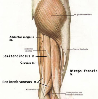
Three long muscles make up this group. The biceps femoris, which is situated along the postero-lateral side of thigh; the semimembranosus, which lies along the medial border and posterior side of thigh; the semitendinosus, which is narrower than the other two muscles of the group, being situated along and over the semimembranosus. All three of them arise from the tuberosity of ischium (of pelvis) and are inserted into the superior portion of tibia. Action: they flex the leg and extend the thigh. They are innervated by the sciatic, tibial, and peroneal nerve. They are supplied by the medial circumflex, perforating, and the popliteal artery, which are branches of the femoral artery.
Below, anterior group of muscles of thigh. The sartorius and the quadriceps are visible in this picture of right thigh. The vastus intermedius cannot be seen as it is located under the rectus femoris. However, three muscles of the medial group can be observed (adductor longus, adductor magnus, and gracilis).
Below, medial group of muscles. View of medial aspect of right human thigh. The adductor brevis is not visible as it lies beneath the gracilis and the adductor longus.
Muscles of thigh. Posterior group: semitendinosus, semimembranosus, and biceps femoris. The adductor magnus and gracilis belong to the medial group.


0 $type={blogger}:
Post a Comment