Chondroglossus Muscle

The chondroglossus muscle is one of the four extrinsic muscles of human tongue. It arises from the lesser horn of the hyoid bone by a small muscular slip. Then its fibers travel up obliquely and anteriorly between the muscular fibers of the hyoglossus as they get mixed up. It is inserted into the dorsum of the tongue, interlacing with the fibers of the superior longitudinal muscle of tongue.

Action

The chondroglossus pulls the tongue downwards and backwards, working together with the hyoglossus.

Innervation and Blood Supply

It is innervated by the hypoglossus nerve (CN XII) and it receives oxygenated blood from small collateral branches of the lingual artery.

Below, right side of human face, exhibiting the extrinsic muscle of tongue. To expose the chondroglossus, the hyoglossus has been cut off.


Genioglossus Muscle

The genioglossus muscle is located at the base of the tongue on both side of its septum. Triangular in shape, it is one of the extrinsic muscles of this organ. It originates from the mental spine of the mandible (called symphysis), fanning out as it extends backwards to insert into the lingual fascia and the hyoid bone.

Action

The genioglossus protrudes (sticks out) and depresses the tongue, pulling it forwards and backwards. Not only does it contribute in the motor articulation of this organ to produce speech, but it is also one of the muscles involved in deglutition (swallowing) of food, a vital reflex action whose center is located in the medulla oblongata.

Innervation and Blood Supply

The genioglossus muscle is innervated by the hypoglossal nerve and is supplied by collateral branches of the lingual artery, which arises from the external carotid artery.

Below, right side of the human face, showing the genioglossus and other extrinsic muscles of the tongue.


Hyoglossus Muscle

The hyoglossus muscle is a paired extrinsic muscle of the human tongue. Flat and rectangular in shape, it is located laterally of the genioglossus, at the base and under this organ of speech.

The hyoglossus originates from the superior border of the body and the greater horn of the hyoid bone. Then its fibers travel upwards and anteriorly towards the lateral margins of the root and body of the tongue, where they run between the styloglossus and  inferior longitudinal muscle, some reaching the tip of the tongue.

Action

The hyoglossus muscle pulls the tongue backwards and downwards, especially when we swallow our food.

Innervation and Blood Supply

It is innervated by the hypoglossal nerve (XII CN) and it receives oxygenated blood from the suprahyoid artery (a branch of the lingual a.) and secondary branches of facial artery, both of which arise from the external carotid artery.

Below, two pictures of human tongue and face, showing the hyoglossus and other extrinsic muscles of this organ.


Styloglossus Muscle

The styloglossus muscle is one of the four extrinsic muscles of the human tongue. It arises from the styloid process and the stylohyoid ligament. Then it travels down obliquely and anteriorly, between the stylohyoid muscle and the pharynx. As it descends, it adjoins the lateral surface of root of tongue and the upper lateral surface of the hyoglossus muscle.

The thicker upper bundle of the styloglossus runs along the lateral margin of the tongue, all the way forward to its tip. Meanwhile, its thinner lower bundle penetrates the hyoglossus muscle and passes downward at the posterior part of tongue to interlace with the tendinous band of its contralateral muscle. This happens on both lateral sides of tongue, as it is a paired muscle.

Action/Function

The styloglossus muscle pulls the tongue backwards and upwards.

Innervation and blood supply 

It is innervated by the hypoglossal nerve and it is supplied with oxygen-rich blood by the sublingual artery, which is a small branch of the lingual.

Down below, illustrative image showing the location of styloglossus in relation to the other muscles of tongue.


Muscles Involved in Jumping

There are several muscles involved in jumping. They are among the largest, longest, and most powerful skeletal muscles of the human body. Also involved in sprinting, they are located in the pelvis, thigh, and leg regions. They are often used by soccer and basketball players when they leap or spring. According to their importance when performing this action, they are:

1) triceps surae, which is a muscle group made up of the gastrocnemius, which consists of two heads, and the soleus muscle, both sharing the same tendon: the Achilles tendon, that is inserted into the calcaneum bone. Action: plantar flexion, raising the body upwards when we stand on tiptoe, and off the ground at the moment of jumping. Innervation: tibial nerve (from sacral plexus);

2) quadriceps femoris, which is a group made up of four muscles (rectus femoris, vastus lateralis, vastus medialis and vastus intermedius); action: they prop up the full weight of the body when we flex the knee just before jumping high, extending the leg at the knee-joint at the moment of jumping. Innervation: femoral nerve (from lumbar plexus);

3) gluteus maximus, which arises from the back of pelvis (ilium) and is inserted in posterior side of proximal portion of femur; action: it extends the thigh, pulling the femur backwards when running and jumping. Innervation: inferior gluteal nerve (from sacral plexus);

4) posterior group of thigh muscles: semitendinosus, semimembranosus, and biceps femoris. Action: they extend the thigh at the hip-joint, flexing the leg at the knee-joint. Innervation: branches of the sciatic nerve (from L4-S3 of lumbo-sacral plexus).

Below, the triceps surae group: the gastrocnemius extending superficialis over the soleus.


Triceps Surae Muscle

The triceps surae muscle is located on the posterior side of leg, allowing us to walk on tiptoe and jump. Broad and powerful, it is composed of two well defined portions: the gastrocnemius and the soleus muscle, both being united into one by the Achilles tendon, which is inserted into the calcaneus (heel bone).

It is called ‘triceps’ because the former consists of two strong heads, which protrudes sideways in athletes, plus the latter, which lies underneath it. Lying on top of the soleus, both heads of the gastrocnemius originates from the distal end of femur, while the soleus arises from the upper portion of fibula and the shaft of tibia.

Action/function

The triceps surae is the muscle of athletes and ballet dancers as it accomplishes plantar flexion of the foot, pulling its tip downwards (when we are sitting) and raising the heel off the ground (when we are standing), allowing us to jump or walk in tiptoe.

Nerve Supply

It is innervated by the medial popliteal nerve (L4, L5; S1, S2), which originates from the sciatic.

Blood Supply

The triceps surae muscle receives oxygenated blood from the posterior tibial and peroneal artery.

Below, an schematic drawing the muscles of posterior aspect of leg, showing the gastrocnemius and the soleus muscle.


Gastrocnemius Muscle

The gastrocnemius is a strong muscle lying on the superficial and posterior aspect of leg. It consists of two heads, a medial and a lateral head. The medial head is stronger, arising from the popliteal surface of the femur. The lateral portion, on the other hand, originates from a point located right above the lateral condyle of the same bone. At their origins, both heads constitute the inferior border of the popliteal fossa.

The gastrocnemius muscle runs down along the posterior side of the leg, over the soleus. In the middle of leg, both heads unite into one muscle, which tapers into a strong, thick tendon in the lower third of the leg; it is the Achilles tendon, with which it shares with the soleus muscle. This sturdy tendon is inserted into the posterior surface of the calcaneus (heel bone).

Action/function

Along with the soleus, it performs two important functions: 1) it performs plantar flexion of the foot, pointing the toes downwards, raising the heel off the ground as when we walk on tiptoe; 2) it contributes to the biceps femoris muscle in the flexion of the leg backwards and upwards at the knee-joint.

Innervation

The gastrocnemius muscle is supplied by the tibial nerve, which is a branch of the sciatic nerve.

Blood Supply

It receives oxygen-rich blood from the posterior tibial and peroneal artery.

Below, image of posterior side of right leg, showing its superficial muscles: the gastrocnemius and the soleus, with the Achilles tendon, which joins them together at the heel.


Soleus Muscle

The soleus muscle is a flat skeletal muscle which is located on the posterior side of leg. It lies under the gastrocnemius and it can only be partially noticed at the medial and lateral aspect of leg.

The soleus muscle arises from the head and upper third of the shaft of fibula, and also from soleal line and the middle third of the shaft of tibia. Then this muscle stretches down towards the heel as it becomes narrower.

In the lower third portion of leg, the soleus muscle fuses together with the gastrocnemius, ending up in a powerful thick tendon; the Achilles’ tendon, which is inserted into the posterior surface of the calcaneus of tarsus (heel bone).

Action/function

The soleus muscle works in unison with the gastrocnemius, performing plantar flexion of the foot as they pull its tip downwards (like a ballet dancer). When one is standing, it raises the heel off the ground.

Innervation

This leg muscle is innervated by the tibial nerve (L4, L5; S1, S2), which arises from the sciatic.

Blood Supply

The soleus is supplied by branches of the posterior tibial and peroneal artery.

Below, an anatomical drawing which shows both the gastrocnemius and the soleus muscle beneath it.


Popliteal Fossa

The popliteal fossa (fossa poplitea, in Latin) is an anatomical region located on the posterior surface of the knee-joint region. It is rhomboid in shape. Superiorly and medially, it is bounded by the semimembranosus and semitendinosus muscle. Superiorly and laterally, it is limited by the biceps femoris muscle. Inferiorly, it is confined by both heads of the gastronemius muscle and the plantaris muscle. The floor of the fossa is constituted by the popliteal surface of the distal end of femur and the posterior surface of the knee-joint capsule.

The region of the popliteal fossa is crisscrossed by a network of arteries that supply the different muscles and bones of the distal portion of thigh and the leg. These are the branches of the popliteal artery, which in turn is the continuation of the femoral artery. It is innervated by the popliteal nerve as well as by the tibial and the common peroneal nerve, all of them being branches of the sciatic nerve.

Below, diagram showing the popliteal fossa region, with the different muscles, arteries and nerves.


Muscles of Thigh

The muscles of thigh are the longest and most powerful muscles of the human body. They consume a large amount of calories (glucose/fatty acid) when we run. Not only are the muscles of thigh long but some of them are also broad and spindle-shaped. The sartorius is the longest and narrowest, while the adductor magnum is the broadest, with the quadriceps femoris being the largest. They are innervated by branches of the femoral, the sciatic, the tibial, and the peroneal nerve and they are supplied by the femoral artery and its branches. Some of them arise from the different anatomical parts of pelvis, but others from the upper third portion of femur, inserting into the tibia.

Anatomically, they are classified into three groups: 1) anterior group; 2) medial group; 3) posterior group.

Anterior Group

The muscles of this group are located on the anterior aspect of thigh. It is formed by the sartorius, which is the longest muscle of the body and the most anterior of the thigh, arising from the anterior superior iliac crest and inserting into the tubercle of tibia (tibial tuberosity); and the quadriceps femoris, which is composed in turn of four muscular heads: the rectus femoris, which lies anteriorly; the vastus medialis, which is located towards the medial side of thigh; the vastus lateralis, lying towards the lateral aspect of upper limb; and the vastus intermedius, which also lies anteriorly but under the rectus femoris.

Medial Group

There are five muscles in this group as their function is the adduction of the thigh (pulling it inwardly as when we ride a horse); they all lie on the medial (inner) aspect of thigh. It is composed of the gracilis, which is long and flattened; adductor longus, which is also long but rather triangular in shape; the adductor brevis; adductor magnus, which is the broadest of them all; adductor minimus; and pectinius muscle, which is small, flat and rather square. These muscles originate from the pubic border of pelvis and insert into the middle line of femur, and into the tibia. They are innervated by branches of the femoral and obturator nerve and they are supplied by the femoral artery. 

Posterior Group 

Three long muscles make up this group. The biceps femoris, which is situated along the postero-lateral side of thigh; the semimembranosus, which lies along the medial border and posterior side of thigh; the semitendinosus, which is narrower than the other two muscles of the group, being situated along and over the semimembranosus. All three of them arise from the tuberosity of ischium (of pelvis) and are inserted into the superior portion of tibia. Action: they flex the leg and extend the thigh. They are innervated by the sciatic, tibial, and peroneal nerve. They are supplied by the medial circumflex, perforating, and the popliteal artery, which are branches of the femoral artery.

Below, anterior group of muscles of thigh. The sartorius and the quadriceps are visible in this picture of right thigh. The vastus intermedius cannot be seen as it is located under the rectus femoris. However, three muscles of the medial group can be observed (adductor longus, adductor magnus, and gracilis).


Below, medial group of muscles. View of medial aspect of right human thigh. The adductor brevis is not visible as it lies beneath the gracilis and the adductor longus.


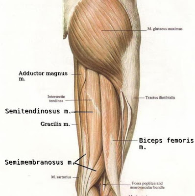
Muscles of thigh. Posterior group: semitendinosus, semimembranosus, and biceps femoris. The adductor magnus and gracilis belong to the medial group.


Quadriceps Femoris

The quadriceps femoris is the strong extensor muscle which lies on the anterolateral aspect of thigh. It is made up of four smaller muscles; they are the rectus femoris, the vastus lateralis, the vastus medialis, and the vastus intermedius, forming a large dense mass covering the front and sides of the femur.

Each of the heads of these muscles arises independently, but as they reach the knee-joint the form one tendon. This means that the individual tendons of these four muscles that constitute the quadriceps fuse into a common tendon at the distal part of the thigh and inserts into the patella and the tibial tuberosity.

The rectus femoris occupies the anterior surface of thigh, arising from the anterior inferior iliac spine by a fine tendon. The vastus lateralis lies on the anterolateral aspect of thigh, originating from the greater trochanter and the lateral lip of linea aspera of femur. The vastus medialis is located on the anteromedial side of thigh, taking origin also from the lateral lip of linea aspera of femur. The vastus intermedius lies on the anterior side of thigh, beneath the rectus femoris muscle, arising from the anterior surface of femur.

Action

Contraction of all the heads of the quadriceps femoris muscle extends the leg at the knee-joint, while the rectus femoris alone flexes the hip-joint. All of them contract when we rise up from a sitting position, and when we run and do sit-ups in the gym.

Innervation

This group of four muscles is innervated by the femoral nerve (from L2, L3 and L4).

Blood Supply

The quadriceps femoris is supplied by the lateral circumflex femoral and the profunda femoris artery.

Below, a diagrammatic picture of right thigh, showing three of the four muscles that make up the quadriceps group. The vastus medialis cannot be seen because it lies underneath the rectus femoris muscle.


Posterior Group of Thigh Muscles

The posterior group of thigh muscles is sometimes referred to as the hamstring muscles. It is composed by three long powerful muscles, all of them arising from the ischial tuberosity of pelvis, stretching along the full length of femur. Their upper (proximal) portions are covered by the gluteus maximus. They are:

1- The semimembranosus muscle, which lies on the medial border of the posterior side of thigh. Origin: ischial tuberosity. Insertion: medial condyle of tibia and oblique posterior ligament of knee.

2- The semitendinosus muscle, which lies closer to the medial border, extending over the semimembranosus. Origin: ischial tuberosity. Insertion: tubercle of tibia.

3- The biceps femoris muscle, which extends vertically on the lateral border of posterior side of thigh. Long and flattish, it arises from the ischial tuberosity of pelvis ischium and get inserted into the proximal end of tibia.

Action/function

They extend the thigh at the hip-joint, flexing the leg at the knee, rotating medially and laterally.

Innervation

The posterior group of thigh muscles are innervated by the tibial (L4-L5-S1) and sciatic nerves (S1-S2).

Blood Supply

They receive oxygenated blood from branches of the medial circumflex femoral, perforating, and popliteal artery.

Below, the schematic picture of posterior aspect of right thigh, showing all three muscles of posterior group.

Biceps Femoris Muscle

The biceps femoris is a long muscle lying on the lateral border of posterior aspect of human thigh. Thus, it belongs to the posterior group of thigh muscles, together with the semitendinosus and semimembranosus muscle. It has two heads, a long and a short head, which fuse together to form a single common belly.

The long head of the biceps femoris muscle arises from the ischial tuberosity by a narrow, flat tendon. The short head originates from the lateral lip of linea aspera of the distal half of femur. Then the two heads get together to become a strong muscle belly, which stretches down along the posterior lateral border of thigh, parallel and next to the semitendinosus muscle. The distal portion of the biceps femoris becomes a long narrow tendon, which curves around the lateral epicondyle of femur to be inserted into the head of fibula.

However, some bands of its distal tendon runs almost horizontally to be inserted into the edge of superior articular surface of the tibia. The lower bursa of the biceps femoris muscle is lodged between its tendon and the lateral ligament of the knee.

Action/Function

It extends the thigh at the hip-joint. It also flexes the leg at the knee-joint, rotating it laterally.

Innervation

The long head of the biceps femoris in innervated by branches of the tibial and sciatic nerve (S1, S2). The short head receives branches from the common peroneal nerve (L4, L5, S1).

Blood Supply

It receives oxygenated blood from the medial circumflex femoral, perforating, and popliteal artery.

Above, a schematic picture of posterior side of right thigh, showing the biceps femoris muscle. You can observe that its proximal (upper) portion is covered by the gluteus maximus.

Semitendinosus Muscle

The semitendinosus muscle belongs to the posterior group of muscles of human thigh. It is long and narrow and stretches along the full length of femur, over the semimembranosus muscle. It lies closer to the medial (inner) border of the posterior aspect of thigh, next and parallel to the biceps femoris mucle. Its proximal end is covered by the gluteus maximus muscle.

The semitendinosus muscle arises from the tuberosity of ischium, traveling downwards along the length of thigh, partially covering the semimembranosus muscle. At its distal extremity, it ends up in a long tendon that curves around the medial femoral epicondyle as it runs down slightly beyond the knee-joint. Then the tendon stretches on to the anteromedial surface of tibia to be inserted into its tuberosity.

Action/Function

The semitendinosus muscle flexes the leg at the knee-joint and it also extends the thigh at the hip joint.

Blood supply

It is supplied by the perforating arteries, which are branches arising from the deep artery of thigh, also known as the deep femoral artery.

Innervation

It is innervated by branches springing from the tibial nerve (L4, L6, S1, S2)

Below, diagrammatic picture that shows the thigh posterior side muscles, which include the semitendinosus, semimembranosus, and biceps femoris muscle.


 The superficial aspect of posterior side of side. The semitendinosus muscle can be perceived as it stands out distintively under the skin in athletic humans, such as soccer and football players.