Environment and Evolution

The environment and evolution of Homo sapiens are intrinsically related to one another. A harsh landscape, with particular climatological circumstances, favored evolution for the better, which was the result of forced adaptation. The disappearance of tropical rain forest and the proliferation of grass and grassy plains during the Miocene and Pliocene boosted the proliferation of ruminants. When these grass-eating quadruped thrived, so did the carnivores and, later, the meat-eating hominid, who was at the beginning a bone breaker that ate the bone marrow of big animals as source of calories.

Idealist people, especially vegans, think that the human evolution took place in an ideal paradise-like environment; a tropical land covered by a jungle full of fruit trees and black soil that spat out potatoes and pumpkin onto the surface of the ground. The bloated minds of utopian people imagine that our ancestors could grab a banana by simply stretching their arm, and if they got tired of banana, he could simply walk a few meters and pick peaches from the peach tree; or he could walk over to the apple tree. Vegans think like that because that is what they do when they go to a supermarket. They just pick the fruit they feel like eating on that particular day, but they never stop to think that all those fruit and vegetable come from different far-flung lands, with different weather patterns, and they are brought to site and concentrated in such great amount by modern means of transportation, and that they are not the product of nature’s generosity but of modern agriculture and industry.

Things were not that easy for our ancestors hundreds of thousands of years ago, for the relation between environment and evolution of Homo sapiens is the story of fighting for survival. There was no tropical fruit or potatoes cropping up out of the earth by magic, for the strife to keep themselves alive took place on the dry grassland of the African savanna and on the cold grassland of the Asian and European steppes and plains. Every discovery of Homo erectus (as well as of the Neanderthal and Cro-Magnon) fossil remains took place in rough environments where the grass grow wild.

Where there is grass growing, there is no jungle. But, on the grassland, there were more important and vital things to eat than fancy fruit, and these things were key to the evolution of our brains. Those vital things are the ruminants (bison, gnu, deer, springbok, goats, sheep, etc) and horses. Homo erectus, the first human, did not have a digestive enzyme that enabled him to break down the coarse grass of the plain, but he did have (as well as today) two powerful enzymes that could digest the four-legged animals that ate the grass: lipase, which breaks down fat, and chymotripsin, which dissolve proteins; these are innate enzymes.

At the beginning, as he lacked the strength and speed of the ferocious feline beasts, he could only wait until a bison or a gnu was hunted by lions as he watched from a distance; his erect posture and stereoscopic vision made of him a sharp observer. Once the big cats ate the entrails and soft meat, the hyenas came over to eat the sinewy muscles and tendons. Then it was man’s turn to chase away the vultures and proceeded to tap the hardest pieces of the carcass to obtain the most delicious and nutritious part of an animal; the bone marrow and the brains.

Although he did not have fangs, serrated teeth, and strong jaws to crack open the long bones and skulls, he had a pair of hands with powerful thumbs opposing the other four fingers which gave him a powerful grab on a stone with which he was able to hammer the bones and skulls open with precision and skills. Then he would discover what an efficient weapon a long bone shard could be when tied to the end of a sturdy stick.

Bone marrow and brains contain high concentration of high quality calories as well as vitamin B12, B6, and B1, vitamin A retinol, zinc, etc. One gram of today’s junk food carbs has only 4 calories, while one gram of saturated fat contains 9 calories, and these are good calories because fat does not raise your sugar level in your bloodstream the way carbs do, but it give you more energy in the form of ketone bodies. Ketone bodies (β-hydroxybutirate and acetoacetate) promotes the bioneogenesis of mitochondria (the production of more mitochondria within a cell, especially within the neurons, accelerating the production of ATP, which means a fast metabolism). This induces the division of the cell and the production of myelin, especially around the cerebral cortex neurons axons.

Below, the grassland of Africa. As on the Asian steppes, there were no bananas, no apples, no pears, no peaches, no flour, no bread, no cereals, no pancakes, no jam, no cakes, no cookies, no beer. There was only tall grass, grove of flat-topped trees in the distance, and wild beasts and ruminants, and slim-wiry, crafty Homo erectuses that strove for survival, using his hands and his good vision.


Artery vs Vein

In the artery vs vein comparison, there are clear functional and structural differences between these two blood vessels. Although they are both parts of the cardiovascular system, there are three important distinctions between them; the function they perform, the type of blood they convey, and the structural feature which one has and the other lacks. Nevertheless, at their extreme ends they are connected by the capillaries, which close the circulatory circuit. At this connection point the arteries and veins are so thin they are called arteriole and venule respectively.

The arteries carry oxygenated blood from the left ventricle of the heart to the rest of the body, with the aorta being the main artery, from which branches arise to supply the different tissues and organs with oxygen and nutrients. The oxygen gets into the blood when we breathe in. They transport the blood under the steady pressure of every systolic contraction of the myocardium; to put it plainly, arteries are able to carry blood thanks to the pumping action of the heart. This is the reason that when we measure somebody’s blood pressure, we do it on an artery.

The veins convey deoxygenated blood from the body tissues and organs back to the heart, to the right atrium, from which it runs into the right ventricle and finally into the lungs. By ‘deoxygenated’ I mean it contains carbon dioxide (CO2), which is expelled from the lungs when we exhale. CO2 is the main byproduct of the cellular respiration when the cells produce ATP (energy). Since they carry the blood back to the heart, that returning blood flow does not depend on the systole (heart ventricles contraction) but on the diastole, which is the heart relaxation; when it relaxes, the myocardium expands outwards as it returns to the normal shape as the right ventricle sucks in the returning blood.

Therefore, when the heart is in diastole, it has a suction action as it draws the blood into the right atrium. This is the opposite to the pumping action of the left ventricle (systole). Since the diastole (relaxation) suction effect is weaker than the systole (contraction), veins are aided by a series of valves, which prevent the deoxygenated blood from flowing back to the capillaries. These valves are located along the lumen of the veins; these valves are not present in arteries.

Below, a picture/diagram of the cardiovascular system, showing the arteries (in red) and veins (in blue).


Medial Inferior Genicular Artery

The medial inferior genicular artery is a blood vessel which lies in the posterior aspect of human knee, right under the medial head of the gastrocnemius muscle. It originates from the medial side of the popliteal artery. Then it runs medially and slightly downwards, curving around the medial periphery of the knee-joint under the medial ligament of knee.

Along its course, the medial inferior genicular gives off secondary branches which anastomose with branches of the medial superior genicular and descending genicular artery, thus being part of the knee vascular network. It supplies the head and tendon of the gastrocnemius, proximal end of tibia, and ligament of knee.

Below, the popliteal artery with its branches, which include the medial inferior genicular artery.


Human Humerus

The human humerus is a long tubular bone of the upper limb (arm). It consists of a shaft and two ends, which are called epiphysis. The upper portion of the shaft is round, while the lower part is trihedral.

The posterior surface of the shaft of humerus features the spiral groove, which lodges the radial nerve. It runs downwards and laterally. Meanwhile, the anterior surface is divided by a poorly defined border into an anteromedial surface and an anterolateral surface, which is the site of origin of the brachialis muscle. The lower half of the anteromedial surface has a nutrient foramen, which leads into a distally running nutrient canal. The deltoid tuberosity is lacated on the anterolateral surface of this bone.

The proximal epiphysis, which is the upper end, is thickened and it features the semispherical head of humerus. The head articulates with the glenoid cavity of scapula and it is separated from the rest of the bone by a shallow annular constriction, which is the anatomical neck.

The greater tuberosity and the lesser tuberosity are located right below the anatomical neck, on the anterolateral surface. They are separated from one another by the intertubercular groove. The greater tuberosity provides attachment to the supraspinatus, infraspinatus, and teres minor muscle, while the lesser tuberosity is the site of attachment for the subscapularis muscle.

The distal epiphysis, or lower end, is flattened in the anteroposterior direction. The distal segment of this bone bears in its lateral part a rounded eminence called the capitulum of the humerus, which is for articulation with the head of the radius bone. The trochlea of humerus is found next to this eminence, which is for articulation with trochlear notch of the ulna.

The distal epiphysis ends up in the lateral and medial epicondyle. The medial epicondyle is larger, with its posterior surface bearing the groove of the ulnar nerve. This groove and the epicondyles are easily palpated (felt with the tips of fingers) under the skin.

Below, the anterior and posterior view of the human humerus (right arm).


Sphenoid Bone

The sphenoid bone is a large butterfly-shaped bone of the skull. Together with the lower part of the occipital bone, it constitutes the biggest part of the base of the human cranium. It is much wider than it is long as its lateral sides stretches out from the center to form the greater wings (right and left) and the lesser wings of bone.

Anteriorly and superiorly, the sphenoid is bounded by the lower border of the frontal bone. Laterally, it is bounded by the temporal, and posteriorly by both the temporal and occipital bone, with which it forms the foramen magnum, which is a large opening for the passage of the spinal cord into the skull. The inferior surface of the orbitofrontal lobe of the brain rests on the lowest part of the greater wing.

Right at the center of the sphenoid, there is a small bone structure called the sella turcica, which is formed by a deep depression known as the hypophyseal fossa; the pituitary gland sits in the sella turcica. The size of the hypophyseal fossa depends on the size of the pituitary. In the greater wing, there are two small openings; one of them is the foramen ovale, which is a passage for nerves and blood vessels; the other the foramen rotundum, which is an small aperture for the maxillary branch of the trigeminal nerve (CN V).

Below, drawing of sphenoid and occipital bone (superior aspect), with names of different parts


Angiogenesis

Angiogenesis is the natural biological process by which new blood vessels are formed. It is a normal process during the development of the fetus and the growth of the body from childhood to adulthood as the need for more blood supply arises. The development of new arterial branches and veins also happens during reparation and replacement of damaged tissue and during the formation of malignant tumor.

Angiogenesis also takes place in the cerebral cortex when new synaptic links are established among neurons during the process of learning new skills, be it intellectual or motor. When there is new synapses in the brain, new support cells, such as oligodendrocytes and astrocytes are formed and the need for more blood supply arises; these cells are the support cells of neuron axons.

Angiogenesis is caused by the development of vascular endothelial cells within a parenchymal tissue as a need to improve the supply of oxygen-rich blood to new areas which are the result of natural growth or development. For example, when an athlete develops muscles when he lifts weights, there is the need for more blood supply for more muscular mass. These squamous endothelial cells multiplication is due to the endothelial growth factor, which is a protein. Endothelial cells make up the single wall of capillaries and they also form the inner wall of arterioles and arteries (tunica intima). As endothelial cells divide, they in turn secrete more growth factor, which stimulate the growth of smooth muscle cells as well. Smooth muscle cells constitute the middle layer of artery wall.

Angiogenesis is very important in the recovery of heart muscle damage after a heart attack caused by coronary artery occlusion. Since the formation of new blood vessels creates important anastomosis (vascular connections/bridges) that prevent damage when an artery or arteriole get occluded or burst. Thus, it is an important biological tool to repair tissue but it also protects both the myocardium and the brain tissue from damage as anastomosis ensure permanent blood supply.

Human Clavicle

The human clavicle is what we commonly call the collarbone. It is a slender, tubular and S-shaped bone. It has a lateral and a medial end. The lateral tip is the acromial end, which articulates with the acromium of scapula. The medial extremity, on the other hand, is called the sternal end, which articulates with the manubrium sterni of sternum.

The acromial end of human clavicle is wider but slightly thinner than the sternal end, which has a sternal articular facet. Looking at it from above, the sternal half of the collarbone makes a forward curve. The inferior surface of the sternal end bears the costal tuberosity, which serves as the site of attachment for the costoclavicular ligament. The inferolateral surface of the lateral end features the acromial articular facet.

The acromial end also bears the conoid tubercle and the trapezoid line, which give attachment to the conoid and trapezoid parts of the coracoclavicular ligament.

Below, the superior and inferior view of right human collarbone.


Thoracic Vertebrae

The thoracic vertebrae are twelve in number. Compared to the cervical vertebrae, they are higher and thicker and they lack the transverse foramen in their transverse process. As the spinal column runs down towards the lumbar vertebrae, their bodies gradually increase in size as they get thicker. There is a concavity on the inferior and superior surface at the base of the vertebral arches; it is the vertebral notch; thus, the two adjacent vertebrae notches form the intervertebral foramen for the spinal nerve roots.

The arches of the thoracic vertebrae form an almost circular vertebral foramina, which is the orifice through which the spinal cord runs; however, it is smaller in diameter than the cervical vertebrae foramina. The transverse process of the thoracic vertebrae extends laterally and obliquely backwards, bearing a small costal facet which articulates with the tubercle of rib. Their spinous processes are long, three-sided, and pointing downwards. Those of the middle thoracic vertebrae fit one over the other, like tiles.

The posterolateral surface of each one of the thoracic verterae bears two facets; the superior costal facet and the inferior costal facet. The inferior facet of one vertebra joins the superior facet of the adjacent distal vertebra (the one below) to give shape to a complete articular facet, which is the site of articulation with the head of the rib. However, the body of the first thoracic vertebra is an exception, for it has a complete articular facet by itself for articulation with the head of the first rib on the superior part of the posterolateral surface, as well as a semifacet for articulation with the head of the second rib on the inferior part.

The tenth vertebra features one superior semifacet on the superior part of the body. Meanwhile, the bodies of the eleventh and twelfth vertebra each bears only one complete costal facet in the middle of each lateral surface.

Below, lateral view of a thoracic vertebra. Every part is labeled

Below, the eighth thoracic vertebra, superior view


Human Scapula

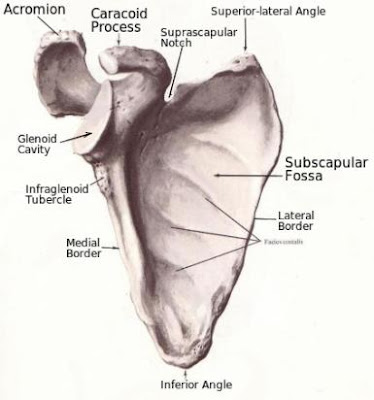
The human scapula, or shoulder blade, is a flat bone of the shoulder girdle region. It lies between the muscles of the back, from the second to the seventh rib. Since it is triangular in shape, three borders and three angles are distinguished in it. The anterior surface is concave and it is called the subscapular fossa, which is occupied by the subscapular muscle.

The posterior or dorsal surface of scapula is divided into two parts by the spine of the scapula. The upper part, which is much smaller, is located immediately above the spine and it is called the supraspinous fossa. The lower larger part occupies the rest of the dorsal surface. They are the site of origin of the supraspinatus and infraspinatus muscle, respectively. Meanwhile, the spine of the scapula, as it extends laterally and upwardly, projects a thick, beak-like protrusion called the acromion. The acromion articulates with the clavicle (collarbone).

The upper border of scapula is thin and features the suprascapular notch. This notch is bridged by the superior transverse ligament of scapula, forming an opening through which the suprascapular nerve goes through. Next to it is the coracoid process, which is a bony protrusion that juts out upwards and then laterally. The coracoid process is the site of origin of two arm muscles; the coracobrachialis and the short head of biceps brachii muscle. The rest of the upper border of scapula is thinner, ending in the superior medial angle, which is also thin.

The superior lateral angle is thick. Right below this angle, the scapula bears a thickened concave surface; this is the glenoid cavity, for articulation with the articular surface of the head of humerus. This union is the shoulder joint. The supraglenoid tubercle is located above the superior margin of the glenoid cavity, while the infraglenoid tubercle is found right below the glenoid cavity. The supraglenoid tubercle is the site of attachment for the tendon of long head of the biceps brachii muscle, while the infraglenoid tubercle is site of attachment of the long tendon of triceps.

Below, anterior view of human scapula.


Below, dorsal view of shoulder blade.


Shoulder Girdle Bones

The shoulder girdle bones are three in number. The scapula, the clavicle (collarbone), and the humerus. They meet at the superior, lateral corner of trunk to form two joints: the shoulder joint and the acromioclavicular joint; the former is the union of the head of humerus with the superior lateral corner of scapula; the latter is the union of collarbone with the acromion of scapula. All three bones are united at this angle by strong ligaments and cartilage.

The humerus and clavicle are longitudinal bones, but the scapula is rather flat and irregular in shape. It projects laterally and upwardly two longitudinal bony protrusions; the acromion and the coracoid process, which resembles a crow's beak. The short head of biceps brachii, the strongest muscle of arm, and the coracobrachialis muscle are attached to the coracoid process.

But the scapula also features on its superior lateral aspect a concave depression; it is the glenoid cavity, into which the head of humerus fits. This is called the shoulder joint. Meanwhile, the lateral end of the clavicle articulates with the acromion of scapula; this is called the acromioclavicular joint.

Below, a shoulder girdle radiograph shows the bones of this portion of the human body. 1- first rib; 2- medial border of scapula; 3- intercostal space; 4- lateral border of scapula; 5- scapula; 6- glenoid cavity of scapula; 7- humerus; 8- greater tuberosity of humerus; 9- anatomical neck; 10- acromion; 11- coracoid process of scapula; 12- clavicle; 13- spine of scapula; 14- superior border of scapula; 15- head of humerus; 16- surgical head.


Human Pelvis

The human pelvis is the wide bone structure at the lower end of the trunk. It supports and contains the last portion of the digestive tract and the urogenital system. Large muscles of the thigh and hip are anchored to it. It articulates with the head of femur at the acetabulum cavity. 

The shape and dimensions of pelvis depend on the sexual differences and individual body size. The average width of female pelvis is 27 cm, while it is 24 cm in the male; this is called the intercristal diameter, extending from the lateral iliac crest of one hip bone to the other of the opposite side. The frontoposterior diameter and pelvic cavity are also wider in female by one and half centimeter.

Anatomical Description

The human pelvis is composed of the two hip bones, the sacrum, and the coccyx. They are all joined together into one anatomical unit by cartilage, ligaments, and joints. The widest part of each one of the hip bones is called the ilium. The upper edge of the ilium is the iliac crest, which runs from back to the frontolateral portion of the pelvis, curving around the side. On the lateral side, the hip bone features a concave cavity; the acetabulum, in which the head of femur fits snugly.

The foremost frontal portion of the hip bone is very narrow compared to the rest and is below the iliac crest; it is called the pubis. The pubis of each hip bone is joined to the one of the opposite side at a medial line by the pubic symphysis, which is a fixed joint made of cartilage. Meanwhile, the bone arch at the bottom and rear portion of the hip bone is called the ischium, which forms an opening referred to as the foramen obturatum. Powerful muscles of the medial side of thigh are attached to the ischium, such as the adductor magnus.

As a whole, the human pelvis can be divided into the greater pelvis and the lesser pelvis. The greater pelvis is bounded on the sides by the wing of the ilium, and posteriorly by the lower lumbar vertebra and the base of the sacrum. Its inferior boundary consists of the crest of the pubis and the frontolateral portion of crest of ilium; this inferior boundary is called the arcuate line of the pelvis.

The lesser pelvis, on the other hand, is located below the arcuate line. Its lateral walls are formed by the lower part of the bodies of the ilia and ischia, the posterior wall by the sacrum and coccyx, and the anterior wall by the two pubic bones.

Below, the female human pelvis, with its different parts labeled. It is wider than the male's.


Coccyx

The coccyx is the distal end of the spinal column in human beings. It is the rudimentary tail of the mammals. Being the continuation of the sacrum, it curves inwardly as it tapers to a point. It is composed of four or five coccygeal vertebrae, which fuse together into one bone structure in adults.

The coccyx has the shape of an inverted pyramid, with its base facing upwards and the apex downwards. The vertebrae that form it are deprived of transverse process and spinous process. They have only bodies. On each side of the first coccygeal vertebra are the remnants of the superior articular processes in the form of small projections, which are called the coccygeal cornua. These extend upward to articulate with the sacral cornua.

The superior surface of the coccyx has a slight concave area, which articulates with the apex of the sacrum by means of the sacrococcygeal joint.

Below, anterior (A) and posterior (B) view of the coccyx.


Cervical Vertebrae

The cervical vertebrae are the seven uppermost bone pieces of the vertebral column. They are located immediately below the skull, with each one of them being separated from the other by a disc of cartilage. They are: C1, C2, C3, C4, C5, C6, C7. The most superior one is called atlas (C1), connecting the base of the skull with the vertebral column. The second cervical vertebra, on the other hand, is called the axis (C2).

From C3 to C7, a cervical vertebra consists of an anterior body; a posterior spinous process, which juts out backwards, ending in a point; two lateral transverse processes; and two inferior articular processes. The vertebral foramen, through which the spinal cord runs, is bounded and contained by these bone processes. The lateral transverse processes of the cervical vertebrae also contain a smaller opening called transverse foramen, through which the vertebral arteries runs up into the head. The distal cervical vertebra articulates with the proximal thoracic vertebra, which is taller and thicker.

Below, a radiograph of the neck region (lateral view). It shows the seven cervical vertebrae. 1- atlas; 2- the axis; 3- transverse process; 4- spinous process; 5- articular process.

Atlas and axis (first and second vertebrae) articulating together (posterior view)


Second cervical vertebra’s lateral view


Second Cervical Vertebra

The axis is the second cervical vertebra. It has an odontoid process, or dens, which is a tooth-like protrusion, lined with cartilage, which articulates with the atlas on top. The atlas, together with the skull turns about the odontoid process as if it were an axis, hence the name. The anterior aspect of the odontoid process bears an anterior articular facet for articulation with the facet for the dens of the first cervical vertebra. Meanwhile, the posterior side of the axis dens has a posterior articular facet, on which the transverse ligament of the atlas adjoin.

At the base of the transverse process on both lateral aspects, the second cervical vertebra bears a superior articular facet, which is for the articulation with the inferior articular facet of first cervical vertebra (atlas). Like the other vertebrae, the axis has a transverse foramen in each transverse process. In the cervical portion of vertebral column, this orifice is for the vertebral artery and vein. On the posterior aspect of its body, it has a short spinous process; the rectus capitis posterior major and the obliquus capitis inferior muscle are attached to the spinous process of the axis.

Below, lateral view of second cervical vertebra, with their different parts


Femur

The femur is the longest and thickest bone in the human skeleton. It is tubular, containing abundant marrow. It is covered and moved by the longest, largest and most powerful muscles in the body, such as the sartorius, quadriceps, and adductor magnus muscle. It is commonly known as the thigh bone.

Anatomical Description

The femur is composed of a body (shaft), and two ends: the proximal end and the distal end. The shaft of femur (corpus femoris) is cylindrical, slightly twisted about its axis, and curved forwards. Its anterior side is smooth, while its posterior surface features a rough linea aspera, which is a long ridge of bone that serves as site of attachment to muscles.

The linea aspera is divided into two lips, the lateral and the medial lip. The lateral lip is the continuation of the gluteal tuberosity. However, in the lower third portion of the femur, it runs down laterally as it extends to the lateral condyle. The medial lip, on the other hand, runs down the length of femur but it swerves out medially in the lower third portion of bone as it curves to the medial condyle.

The proximal, or upper, end has two rough protrusions at the junction with the shaft. These protrusions are the greater and the lesser trochanter. The greater trochanter juts out upwards and backwards, making up the lateral part of the proximal end of femur. Meanwhile the lesser trochanter is located on the posteromedial surface of proximal end of the bone. The intertrochanteric line extends between the greater and the lesser trochanter on the anterior aspect of upper end of femur, while the intertrochanteric crest runs on the posterior side between the these two protrusions.

The neck of the femur arises from the medial aspect of its proximal end, between the greater and lesser trochanter. Then it projects upwards and medially, ending up in a spherical portion, which is called the head of femur (caput femoris). The neck is slightly flattened on its frontal plane. The head has a small rough pit, which is called the fossa of the head of femur, where the ligament of the head is attached. The head fits in the acetabulum, which is a cup-shaped cavity on the lateral surface of hipbone (pelvis). They articulate with one another.

The distal or lower end of femur is thick and wide, terminating in two condyles; a medial and a lateral conyle. The medial condyle is larger than the lateral one and they are separated from one another by a deep depression called the intercondylar fossa where the cruciate ligaments are attached. The medial surface of the medial condyle and the lateral surface of the lateral condyle feature a medial epicondyle and a lateral epicondyle respectively, which are small bone projections that serve as attachments for ligaments and tendons.

Below, the anterior side of right femur, with its different parts.

Below, posterior side of right femur. Notice the linea aspera and the lateral and medial lip.


First Cervical Vertebra

The first cervical vertebra is called the atlas. It is the topmost of the seven cervical vertebrae and, therefore, the proximal one of the spinal column. Its vertebral foramen is aligned with the foramen magnum of the base of skull to allow the spinal cord to extend into the cranium as the medulla oblongata.

The first cervical vertebra looks differently from the rest of vertebrae as it lacks the spinous process on its posterior aspect. Rather, it is an irregular ring formed by two arches: the anterior and posterior arch. These are fused to one another by two thick lateral bone masses. On their superior surface, each of these masses bears an oval, concave superior articular facet, which is for the articulation with the occipital bone. Meanwhile, on their inferior surface, they have a flat inferior articular facet, which is for articulating with the superior surface of the second cervical vertebra (axis).

The 1st cervical vertebra anterior arch has an anterior tubercle and small facet for the odontoid process on the posterior surface which articulates with the dens of the axis. The atlas posterior arch has a posterior tubercle, which represents the missing spinous process (spine). Like the rest of the spinal column vertebrae, the atlas has two transverse processes, one on each lateral aspect of it, with an orifice in each one of them; they are for the vertebral arteries, which arise from the subclavian.

Below, the first cervical vertebra (superior aspect).

Below, inferior view of the atlas.


Snouted Cobra

The Snouted Cobra (Naja annulifera) is a large brown snake of the savanna of southeast Africa. Its glands produce venom with powerful neurotoxins, which cause death from respiratory failure. However, it is not as aggressive as other cobras. It is nocturnal, coming out at dusk to pray on rodents and other small animals.

Physical Characteristics

The Snouted Cobra is light brown in color, but it goes through a banded phase, in which it is black, with yellowish cream bands. A distinctive dark throat can be observed in juveniles. The scales are smooth, with 19 rows at mid body and the ventral side has 175 rows. The head is broad, and the eye pupils are round. It is characterized by having a row of scales between the eye and upper labials.

The female of the Naja annulifera lays 20 eggs in Summer. When it feels threatened, it spreads its broad hood and is always ready to bite. However, it does not spit, as other species do. It inhabits the dry and moist savanna of south and southeast Africa and the woodland, between sea level and the 1,600 m of altitude.

Below, the Snouted Cobra, in its banded phase.

The adult of the Naja annulifera, which is yellowish brown in color.


Cervical Plexus

The cervical plexus is a network of spinal nerve fibers that supply the region of the neck. It consists of fibers from the ventral rami of nerves from C1 to C5. Their branches innervate the skin of the neck and head, as well as the diaphragm and some muscles of neck. The cervical plexus gives off six important nerves:

1) the lesser occipital nerve, which supply the skin of lateral scalp;

2) the greater auricular nerve, which innervates the skin around the external ear;

3) the transverse cervical nerve (also called superficial cervical N.), supplying the skin of ventral and lateral neck;

4) the ansa cervicalis, which is a nerve loop that gives off branches to supply the omohyoid, sternohyoid, and the sternothyroid muscle;

5) the supraclavicular nerve, which innervates the skin of shoulder and central chest;

6) the phrenic nerve, which innervates the diaphragm. The origin of this nerve is found in grey nuclei located in the medulla oblongata and pons.


Elapidae

Elapidae, or elapids, is a taxonomical family of snakes, most of which are very dangerous for humans. They have immovable fangs located at the front of the upper jaw. Their bites are of medical significance as their glands produce powerful neurotoxic venom, which can cause death quickly if untreated.

Genera and Species

The family Elapidae include around 50 genera and 375 species, which are distributed world-wide in tropical regions. They comprise the mambas (Dendroaspis), which are found in Africa, the cobras, the King Cobra (Ophiophagus hannah), the coral snakes, the Australian brown snakes and taipans (Oxyuranus), sea snakes (Hydrophiinae), kraits (Bungarus), and African garter snakes. All African land elapids belong in turn to the sub-family Elapinae. The sub-family Hydrophiinae consist of the Yellow-bellied sea snake and the Australian terrestrial elapids.

Physical Characteristics

The great majority of elapids snakes have long, slender body. Their scales are smooth and usually smaller in size than those of the family Viperidae. They have round eye pupils and their heads are covered by very large scales or shields, being rather elongated and not triangular, as is the case with the viper snakes. Some of them have hood, which they spread out when they feel threatened. Many of them are burrowers, some live on grassy plains, such as the African savanna, others are arboreal. Their females are oviparous, meaning that they lay eggs. Most of them are diurnal (active during the day).

Below, a famous member of the family Elapidae; a cobra, one species from Africa in this case.


Body Tissue Repair

The body tissue repair begins right after the acute inflammatory reaction, which is the result of tissue damage. It is the replacement of dead cells by new viable ones. These new cells may originate from the damaged parenchyma (an organ inherent cellular structure) or from connective tissue in the damaged area.

But parenchymal regeneration is very slow, since the cells that work together to perform the organ main function are stable and it takes a long time for them to multiply, as is the case with hepatocytes, the cells that constitute the liver. When this organ is damaged by either a viral infection or alcohol, it is the organ connective tissue cells that does the repair as they multiply much faster than the parenchymal cells. However, body tissue repair through proliferation of connective tissue is not good, especially for organs such as the liver, lungs, and the heart.

Unfortunately, when the liver is repeatedly wounded by a chronic disease or alcoholism, the liver parenchymal cells (hepatocytes) do not have time to undertake the repair, naturally regenerating, and are overtaken by massive connective tissue cells proliferation; and overtime, the organ become fibrous and rigid, rendering the organ unable to perform its functions. Thus, fibrosis is due to connective tissue repair. Fibrosis in the liver is cirrhosis.

Black Mamba

The Black Mamba (Dendroaspis polylepis) is a long, slender snake which inhabits the different regions of sub-Saharan Africa. It is the most feared snake in that continent. According to recorded hospital statistics, there are an average of 2,500 Black Mamba's bites per year, with 30 deaths, in Africa.

It belongs to the Elapidae family of snakes, like the cobras. Thus, its glands secretes a potent neurotoxic venom. It can bite any part of the victim's body because it has the fangs located well forward on its upper jaw. It is diurnal and feeds mainly on rats and other rodents. It is usually found in the savannas and other grassy plains.

Physical Description

The Black Mamba is dark grey or olive in color, with the belly being creamy or pale green. The upper portion of the body is often speckled with black blotches, especially on the flanks. The adult can measure up to 3.50 m long, but the average length is 2.50 m. It has smooth scales, with 23-25 rows at mid body. Rows of ventral scales are between 239 and 281.

The head is elongated and 'coffin-shaped'. It has round pupils and a pronounced brow ridge. The inside of the mouth is dark bluish or black. Although it is not really black, it is called Black Mamba due to the black mouth lining. The female lays a clutch of 12-18 large eggs each year, with the hatchlings measuring between 45 and 60 cm in length.

Habitat and Distribution

The Dendroaspis polylepis is usually encountered in either the moist and dry savanna areas. However, it can also be found in the coastal bush and the woodland. Although it is usually found in the southern third portion of Africa, it is widespread in this continent. Since this species is widespread, its presence was also reported in the southern border of the Sahara desert. An experienced snake collector in Burkina Faso was bitten three times and killed by a Black Mamba; he had never encountered one before and did not think it was aggressive. This snake moves fast, very fast, and one has to be very careful.

Below, a photo of a Black Mamba taken in the woodland of South Africa.


Lateral Superior Genicular Artery

The lateral superior genicular artery is one of several blood vessels supplying the knee. It arises from the lateral side of popliteal artery. First, it extends laterally, running under the distal portion of the biceps femoris muscle. Then it travels anteriorly as it curves around and above the lateral condyle of the femur bone, hugging it.

When it has reached the frontal aspect of knee, the lateral superior genicular artery divides into several smaller branches, just above the patella. These smaller branches anastomose with other arterial branches to become part of the knee-joint vascular network.

The lateral superior genicular artery supplies the vastus lateralis muscle and the lower part of femur, forming an anastomotic arch across the front of this bone with the highest genicular and the medial inferior genicular arteries.

Below, image of arteries that supply the knee, showing the femoral, the descending genicular, the popliteal, and the lateral superior genicular artery. It is an anterior (frontal) aspect drawing, as you can see the popliteal running down behind the knee-joint.


Artery

An artery is a blood vessel which carries oxygenated blood from the heart left ventricle to the rest of the body. It supplies every tissue and organ with oxygen and nutrients. Its smooth muscle, which forms its middle layer, makes it elastic as its walls can expand outwards when blood pressure increases. It has to be like this, since it must withstand the pressure exerted by the pump (heart), when the ventricular muscle of the heart suddenly contracts during the systole to send blood to every part of the body. Thus, when blood pressure is measured, it is always done on an artery, never on a vein. In anatomical drawings and diagrams, arteries are red colored, while veins are light blue.

The main artery of the body is the aorta, which arises from the left ventricle, ascending and forming an arch, which gives off important branches that supply the head and the upper limbs. Then it runs down parallel to the spinal column as it sends secondary arteries that supply the internal organs. At the base of the abdomen, the aorta divides into two important branches, which are the common iliac arteries. The main artery of the lower limb (thigh and leg) is the femoral artery, while the main artery of the upper limb (arm) is the brachial artery. Meanwhile the head, which include the brain, is supplied by the common carotid and basilar artery.

As it extends throughout the body, the artery keeps giving off secondary branches, just like a tree, forming a network, as it gets smaller and smaller in size, until it becomes so thin that it is called arteriole, which can be thinner than a hair. Finally, the arteriole ends up in a capillary, which is the tiniest blood vessel of the circulatory system. In order to efficiently supply an organ, an artery sends off lateral side branches that link up with another nearby artery; this is called anastomoses, which form a network of arterial blood vessels supplying an organ tissue.

Structure

The artery is made up of three layers of three different tissues: 1) the tunica intima, which is the innermost layer that lines the lumen (opening) of the artery; 2) the tunica media, which is the middle layer; and 3) the tunica adventitia, which is the external coating of the artery. The tunica intima is an endothelial layer, which means it is made up of endothelium (flat epithelial cells that line the inner walls of blood vessels and organs). The tunica media is formed by smooth muscle, which gives the artery elasticity. Meanwhile, the tunica adventitia consists of connective tissue, which give the artery form and consistency.

Innervation

The smooth muscle of tunica media is innervated by the peripheral nervous system (sympathetic and parasympathetic fibers), which means that its constriction and dilation do not depend on our will (cerebral cortex), but on adrenaline and acetylcholine, which are neurotransmitters of the peripheral nervous system.

Below, a diagram of the systemic blood circulation. Arteries are in red, while veins are in blue. The artery coming out of the heart left ventricle is the aorta.


Desert Mountain Adder

The desert mountain adder (Bitis xeropaga) is a venomous snake from the African continent. It is not well known because its natural habitat is the mountain region bordering the lower Orange River, in South Africa and Namibia. Since it belongs to the Viperidae family of serpents, its venom, which can be lethal, contain cytotoxins, which cause blood clots and destroy body tissues.

Physical Description

The desert mountain adder is a medium size snake, with the adult being no longer than 60 cm. It has a big triangular head, with a ridge running above each eye but it has no horns. Its body is greyish-yellowish brown, which is interrupted by multicolor rings composed of dark brown, white, and grey strips. However, its belly is dark grey patterned with dark speckles.

It usually dwells in nook and crannies under rocks and boulders and it never slithers under the sand. It hunts rodents, lizards, and other small animals. The female bears between 4 and 5 young every Summer. Even though it is not aggressive, it could bite you if it is cornered or stepped on.

Below, photo of the desert mountain adder (Bitis xeropaga)


Vastus Lateralis

The vastus lateralis is a long muscle of the anterolateral surface of the human thigh. It is part of the quadriceps femoris group. Its proximal end is covered by the tensor fasciae latae muscle.

The vastus lateralis muscle originates from the greater trochanter, the intertrochanteric line, and the lateral lip of linea aspera of femur. Next, it runs down laterally. Then its distal portion travels anteriorly, ending up in a wide tendon, part of which gets fused with the rectus femoris muscle tendon to be inserted into the lateral border of patella.

Action

The vastus lateralis muscle function is to extend the leg at the knee-joint. In doing so, it contributes to lift up the whole body when it is in a sitting position, as when we stand up from a chair, working together with the other muscles of the quadriceps.

Innervation

For the contraction, the vastus lateralis receives nerve fibers from the femoral nerve (L2-L4).

Blood Supply

It is supplied by branches arising from the profunda femoris artery, which include the lateral circumflex femoral and the perforating arteries.

Below, an anterior view of the right thigh. As you can see, the vastus lateralis muscle is partially covered by tensor fasciae latae


Vastus Intermedius Muscle

The vastus intermedius muscle is one of the four heads of the quadriceps femoris. It lies on the anterior aspect of thigh, between the vastus lateralis and vastus medialis. It is completely covered by the rectus femoris muscle.

The vastus intermedius muscle arises from the intertrochanteric line of head of femur. Then it travels downwards along the femur getting narrower as it goes. When it has reached the middle portion of thigh, it becomes a wide tendon, which fuses with the tendon of the rectus femoris to form the common tendon of the quadriceps femoris muscle.

Action

Extension of leg at the knee joint.

Innervation

This vastus intermedius muscle is innervated by nerve fibers arising from the femoral nerve (L2-L4).

Blood Supply

Like the other muscles of quadriceps, it receives oxygenated blood from the lateral circumflex and profunda femoris artery.

Below, two pictures of vastus intermedius; the first one with the vastus lateralis and the vastus medialis. The other without these muscles.


Rectus Femoris Muscle

The rectus femoris muscle is the longest head of the quadriceps femoris. It occupies the superficial and anterior side of the human thigh. It emerges from the anterior-inferior iliac spine and the superior border of acetabulum (of pelvis). Then it extends anteriorly, all the way downwards, covering the vastus intermedius muscle.

In the distal portion of thigh, the rectus femoris ends up in a narrow yet strong tendon, which fuses with the base and the anterior surface of the patella. Having run beyond the patella, this tendon is called patellar ligament, which is inserted into the tibial tubercle (tuberosity).

Action 

Its main function is the extension of the leg at the knee-joint.

Innervation and Blood Supply

The rectus femoris muscle is innervated by branches of the femoral nerve and it receives oxygenated blood from branches of the profunda femoris artery.


Regions of Abdomen

The regions of abdomen are the different parts into which the human venter (belly) is anatomically divided. The upper part of abdomen has an epigastric region (epigastrium), which is triangular, and two, left and right, hypochondriac regions. In the middle portion of the belly, two lateral abdominal, right and left, regions, and a middle umbilical region. The lower part of the abdomen, called hypogastrium, also has two lateral regions, which are called the inguinal regions (right and left), and a middle pubic region.

 Right and left costal arches are clearly outlined when the abdominal wall is pulled in. The xiphoid process is left in the angle formed by the cartilaginous costal arches, which is called infrasternal angle. The inguinal folds that correspond to the position of the inguinal ligaments are distinctively seen in the lower portion of abdominal wall. When the abdominal muscles are tensed, a groove get formed running vertically along the midline; it is the linea alba abdominis. The umbilicus lies right in the middle of the linea alba, on the level of the articulation between the third and fourth lumbar vertebrae.

The rectus abdominis muscles are outlined to both sides of the midline. The tendinous intersection of these muscles are defined clearly as three or four transverse constrictions. The slips of the external oblique muscle alternating with slips of serratus anterior and longissimus dorsi muscle are outlined on the upper part of the anterolateral portion of thoracic wall.

Below, you can see a schematic drawing of the regions of abdomen and the thorax.


Human Diaphragm

The human diaphragm is an unpaired broad and flat muscle which divides the thorax from the abdominal cavity. Being dome-shaped, it isolates the thoracic viscera, such as the lungs and heart, from the digestive organs (stomach, intestines, liver, and pancreas). It plays a vital role in breathing as it is innervated by the phrenic nerve, whose center is located in the medulla oblongata (see breathing centers).

Both its thoracic and abdominal surface are covered by fasciae, which in turn are covered by connective tissue. A relaxed diaphragm has a beveled spherical convexity, which faces the thoracic cavity with two domes; the right and left. The apex of the dome rises on the medioclavicular line to the level of the fourth intercostal space on the right side, and to the level of the fifth intercostal space on the left. When the diaphragm contracts, its domes flatten out, increasing the thoracic space volume and the capacity of the lungs to expand during the inspiration phase of breathing.

The muscular fibers of the diaphragm arise from the inner border of the outlet of thorax, which can be divided into a sternal, costal, and lumbar portion. The sternal part is the smallest, arising from the posterior surface of the xiphoid and is continuous with the central tendon. The costal portion originates from the inner surface of the bony and cartilaginous parts of the lower six ribs, with its fibers running upwards and inwards, being continuous with the central tendon. The lumbar portion of diaphragm arises from the anterolateral surface of the body of the first three lumbar vertebrae and from the medial and lateral arcuate ligament; at this point, there is the aorta diaphragmatic foramen (opening).

The muscular fibers of the human diaphragm, which runs towards the center, continue as tendons from the central tendon. This area of the diaphragm is trifoliate (having three leaf-like parts) in shape, with one leaf directed forwards and on which the heart lies, and the other two directed to the sides, with the lungs being located there. In the posterior part of the central tendon, to the right of the midline, there is the vena-caval opening for the inferior vena cava. The medial muscular bundles of both crura opens up to form the esophageal opening for both the esophagus and the vagus nerve. There are other smaller orifices in it for the splanchnic nerve and veins.

The lungs and heart lie on the thoracic surface of the diaphragm, while the liver, stomach, and spleen are in contact with its abdominal surface. The pancreas, duodenum, kidneys, and adrenal glands touch areas of the abdominal surface of diaphragm which are not covered by the parietal peritoneum.

Action/Function

The human diaphragm takes part in inspiration (breath in) as it becomes flat upon contraction, increasing the thoracic cavity volume and decreasing its inner pressure. This unbalance between the external atmospheric pressure and the thoracic inner pressure, causes a stream of air to flow into the lungs through the nose to balance it out.

Blood Supply

It is supplied by the phrenic arteries, which originate straight from the thoracic aorta, and by the musculophrenic artery, which springs from the internal thoracic artery.

Innervation

It is innervated by the phrenic nerve, which arises from vertebral roots C3, C4, and C5 as it is part of the cervical plexus.

Below, an anterior aspect of the human diaphragm.