Semimembranosus Muscle

The semimembranosus muscle lies on the medial border of posterior side of the human thigh. Its lateral half is covered by the semitendinosus muscle, leaving a mark in the form of a wide longitudinal groove. With the medial margin of the muscle being free, it stretches along the full length of the femur. It is part of the posterior group of muscles of the thigh.

The semimembranosus muscle originates from the ischial tuberosity of pelvis by a flat, strong tendon. Then it travels downwards vertically along the length of thigh, partially covered by the semitendinosus. It ends up in a distal flat tendon which narrows gradually. This tendon winds around the medial epicondyle as it runs to the anteromedial surface of tibia where it becomes wider and divides into three bands; the first one is inserted into the medial condyle of tibia; the second band fuses with the fascia covering the popliteus muscle; and the third one is inserted into the oblique posterior ligament of knee. 

Function/action 

It extends thigh straight at the hip-joint. It also flexes and rotates leg medially. 

Nerve supply 

The semimembranosus  muscle is innervated by the tibial nerve, which is a major branch of the sciatic nerve. 

Blood supply 

It receives oxygen-rich blood from a branch from the deep femoral artery, which arises from the femoral, and the gluteal arteries, which spring from the internal iliac artery.

Below, posterior aspect of muscles of right thigh, showing the semimembranosus, semitendinosus, and biceps femoris muscle.

The semimembranosus muscle, with the semitendinosus cut away to expose the groove along its lateral half.


Middle Meningeal Artery

The middle meningeal artery arises from the superior side of first part of maxillary artery (mandibular portion), being its largest branch. Then it travels upwards and passes through the foramen spinosum into the cavity of the skull, where it divides into a frontal and a parietal branch. These two main branches extend across the external surface of dura mater in the arterial sulci of the cranial bones. They supply the sulci as well as the temporal, frontal, and parietal areas of dura mater.

Along its course, the middle meningeal artery gives off the following branches: 1) the accessory meningeal artery, which originates from the extracranial part of the middle meningeal to supply the pterygoid muscles, the pharyngotympanic tube, and the muscles of palate; 2) superior tympanic artery, which supplies the mucous membrane of tympanic cavity; 3) superficial petrosal branch, which arises above the foramen spinosum and then stretches laterally and to the back to supply bone tissue.

Below, a picture of the right side of face showing the maxillary and the middle meningeal artery.


Branches of Maxillary Artery

All the branches of maxillary artery carry about 50% of the blood supply to the face and ear. Arising from the external carotid artery, the maxillary has a rather long and tortuous course. For this reason, it can be divided into three portions for a better comprehension.

First Portion (mandibular part)

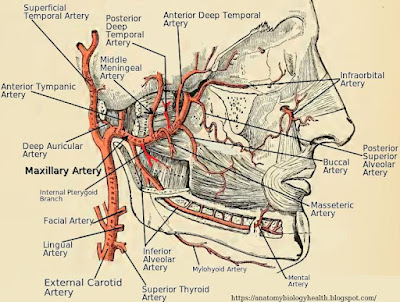
As soon as it springs from the external carotid, the maxillary gives off five branches, with the first four arising from its superior side; 1) the deep auricular artery, which runs upwards to supply the auricular capsule of the mandibular joint, the inferior wall of the external auditory meatus, and the tympanic membrane; 2) the anterior tympanic artery, which travels up and enters the tympanic cavity through the squamotympanic fissure to supply its mucous membrane; 3) the middle meningeal artery, which is its largest branch, running upwards through the foramen spinosum into the skull to supply a large part of the dura mater that covers the frontal part of the brain; 4) the inferior alveolar artery, which is the only one that originates from the inferior side of first portion of maxillary as it travels downwards through the mandibular foramen to supply the chin and lower teeth.

Second Portion (pterygoid part)

The second part of the maxillary gives off several branches which originate both from its superior and inferior side. 1) The posterior deep temporal artery, which arises from its superior aspect and runs upwards to supply the temporal muscle, periosteum, and bone of the temporal fossa; 2) The anterior deep temporal artery, which also originates from the superior side of maxillary and irrigates the temporal muscle 3) the pterygoid branches, originating within the pterygoid bone, with one branch traveling upwards and the other downwards in the oppossite direction, to supply the lateral and medial pterygoid muscle respectively; 4) the messeteric artery, which descends through the mandibular notch to provided the messeter muscle with oxygenated blood; 5) the buccal artery, which also descends to supply the buccinator muscle and the upper portion of cheek.

Third Portion (pterygopalatine part)

The third and final section of the maxillary artery is located in the pterygopalatine fossa. There, several smaller branches arise from it to provide the upper and medial part of face with oxygenated blood. 1) The posterior superior alveolar artery, which originates from its inferior side and travels downwards and obliquely to supply the superior premolar and molar teeth, gingiva (gum), and maxillary sinus; 2) the descending palatine artery, which first gives off the artery of the pterygoid canal, then it divides into the greater and lesser palatine artery that supply the soft and hard palate as well as the gum and bone of the lower and posterior part of maxilla; 3) the infraorbital artery, which travels down to supply the roots of the superior incisors and canine teeth; 4) the sphenopalatine artery, being the terminal branch of the maxillary that runs through the sphenopalatine foramen to supply the posterior part of nasal cavity and the septum.

Below, schematic picture showing the branches of maxillary artery.


Deep Auricular Artery

The deep auricular artery is a deep blood vessel of the side of the face. It originates from the superior side of first part of maxillary artery (mandibular portion), being its first branch. In some individual, it arises together with the anterior tympanic artery, sharing a common trunk.

The deep auricular artery travels upwards and slightly backwards through a portion of parotid gland. Running behind the temporomandibular articulation, it follows an oblique course to penetrate the wall of the external auditory meatus. It gives off collateral branches that supply the articular capsule of the mandibular joint and the inferior wall of the external auditory meatus, while its terminal branches supply the tympanic membrane.

Below, a picture of right aspect of human face, showing the maxillary with all its branches. You can see the deep auricular artery in stronger red color, arising as its first branch.


Infraorbital Artery

The infraorbital artery is a blood vessel of the face which supplies the muscles of the eyeball and the front teeth of maxilla. It arises from the third part of maxillary artery in the pterygopalatine fossa, sharing a common trunk with the posterior superior alveolar artery.

From its point of origin, the infraorbital artery runs upward through the inferior orbital fissure into the eye orbit, fitting into the infraorbital groove (sulcus infraorbitalis). Then it lies in the infraorbital canal as runs. It finally emerges from the infraorbital foramen onto the face where it gives off terminal branches to the tissues of the infraorbital region.

Along its course, the infraorbital artery gives off the following branches: 1) ocular arteries, which supply the inferior rectus and inferior oblique muscle of eyeball; 2) anterior superior alveolar arteries, which bring oxygenated blood to the incisors, canines and the gum of maxilla as well as to its mucous membrane.

Below, a schematic picture of arteries of face, showing the posterior superior alveolar and infraorbital artery originating together as a common trunk from the third portion of maxillary artery. The zygomatic bone is missing so that the point of origin of artery can be seen.


Maxillary Artery

The maxillary artery is one of the three blood vessels which supply the human face. The others are the transverse facial and the facial artery. It has a tortuous course and gives off important deep branches that bring oxygenated blood to the upper portion of face, middle ear, muscles of mastication, teeth and gum of mandible and maxilla, hard palate, nasal cavity, and other deep structures of face.

The maxillary artery arises from the anterior aspect of external carotid artery, at a right angle, at the level of the neck of mandible. At the beginning, it is covered by the parotid gland. Then it runs tortuously and horizontally to the front between the ramus of mandible and the sphenomandibular ligament. Next, it extends between the lateral pterygoid muscle and the temporal muscle. As it runs into the pterygopalatine fossa, it divides at the end into terminal branches.

According to the anatomical topography, the maxillary artery can be divided into three parts, or portions: 1) the mandibular portion (first part), which gives off the deep auricular, anterior tympanic, middle meningeal, and inferior alveolar artery; 2) the pterygoid portion (second part), which sends the posterior deep temporal, masseteric, buccal, and anterior deep temporal artery, as well as two pterygoid branches; 3) pterygopalatine portion (third part), which lies in the pterygopalatine fossa, giving off the posterior superior alveolar, infraorbital, descending palatine, and sphenopalatine artery.

Below, two schematic pictures of the maxillary artery and its branches.


Sphenopalatine Artery

The sphenopalatine artery is a deep blood vessel of the face. It supplies part of the nasal cavity, septum of nose and part of pharynx with oxygenated blood. It arises from the third part of maxillary artery (the pterygopalatine portion), being its terminal branch.

Once it has arisen, the sphenopalatine artery runs upwards, as it goes through the sphenopalatine foramen. It enters the cavity of the nose, where it ramifies to form the following vessels:

1) the supreme pharyngeal artery, which supplies the upper border of pharynx;

2) the posterior lateral nasal arteries, which bring oxygenated blood to the lateral wall of the cavity of nose, the middle and inferior nasal conchae, and the mucous membrane of the frontal and maxillary sinus;

3) the posterior septal nasal branch, which supplies the mucous membrane of the septum of the nose. It anastomoses with the greater palatine and the superior labial artery.

Below, saggital image of right side of nose and oral cavity, showing the origin and branches of the sphenopalatine artery.